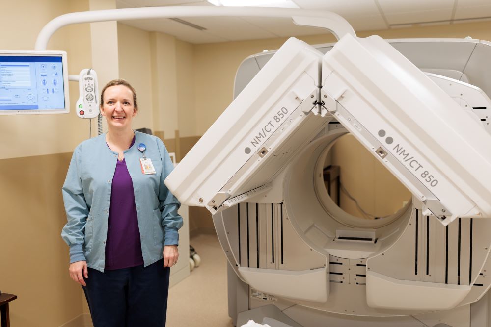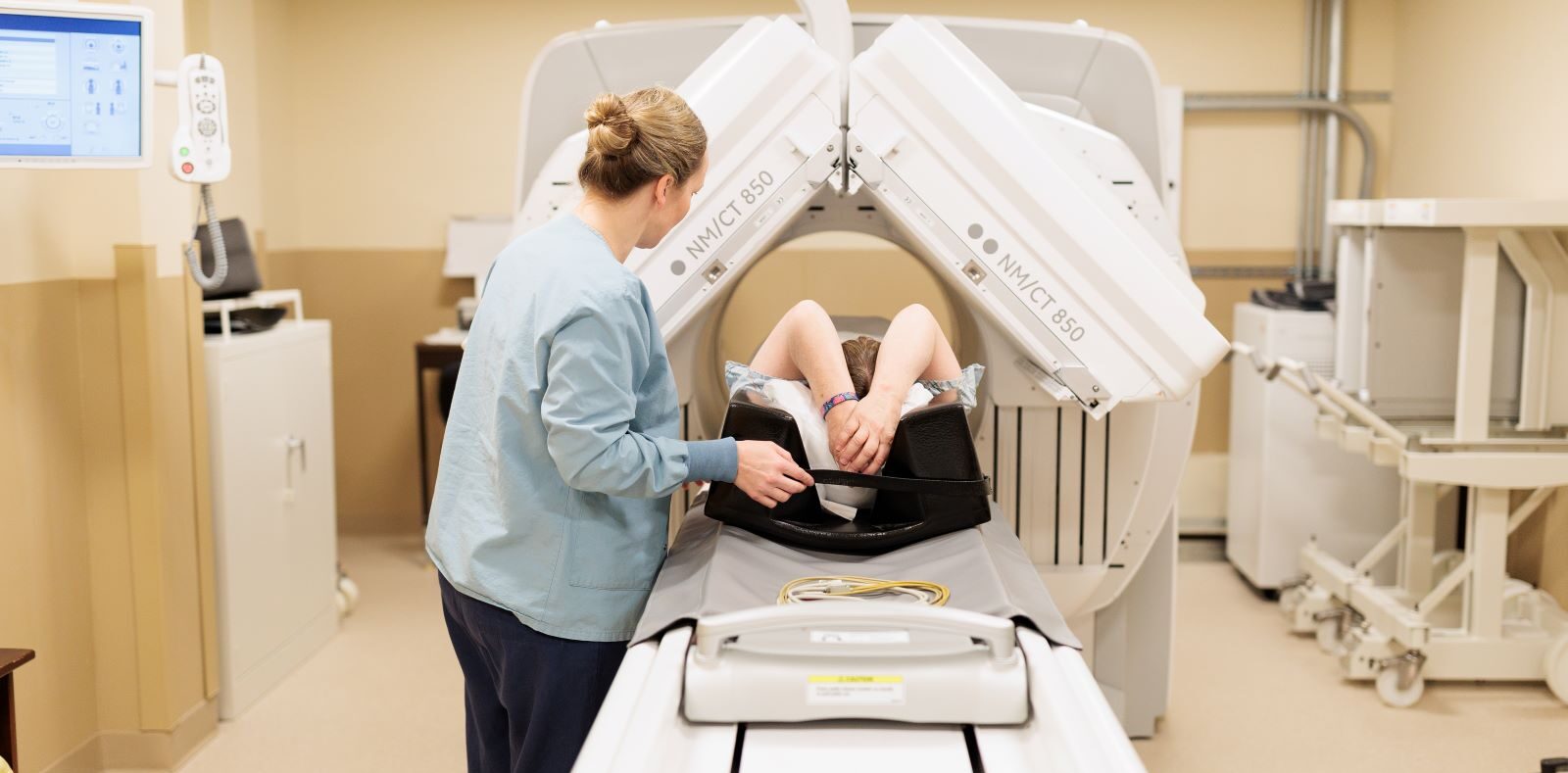
Gifford’s Nuclear Medicine Department offers specialized imaging that shows the function and structure of organs. These images are used to identify problems that routine X-rays, ultrasounds, CT scans and MRIs are unable to diagnose.
Tests commonly performed in our Nuclear Medicine department include:
- Stress testing
- Gastric emptying studies
- Bone scans
- Hepatobiliary studies
- Renal scans
- MUGA (multi-gated acquisition) scans
- Sentinel node scans
- White blood cell scans
- Gastrointestinal bleeding scans
- Electroencephalogram (EEG)
Nuclear stress testing is used to look at the blood flow to the heart muscle and evaluate heart function. This test can diagnose coronary artery disease and is also used to evaluate a patient’s progress after a heart attack or heart surgery.
Hepatobiliary studies are used to evaluate the function of the gallbladder.
MUGA scans are commonly performed for patients who are starting or who have completed cardio toxic chemotherapy. This test can evaluate the left ventricular ejection fraction to determine if the chemotherapy has had a negative effect on the heart muscle.
Bone scans can diagnosis stress fractures, infections and bone metastases.
Electroencephalogram (EEG) is a noninvasive test that measures and records the electrical activity of the brain by placing small electrodes on the patients scalp. This test is most commonly used to diagnose seizure disorders and is read by Gifford’s neurologist.


